
Fig. 8.
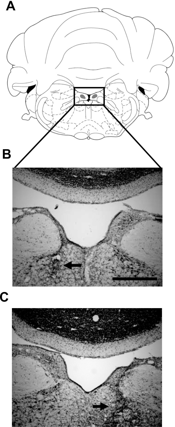
A: section at −10.3 mm relative to bregma reproduced with permission from Paxinos and Watson (1998). B: corresponding section from a rat with complete neurotoxic damage to the right SGN (ipsilateral to the electrode implant; jb25). Note that complete sparing of the left SGN occurred in this animal. Scale bar, 500 μm. C: section from an animal with complete neurotoxic damage to the left SGN (contralateral to the electrode implant; jb28). Note that the right SGN is completely spared. Horizontal arrows in B and C indicate regions where the lesion extended ventrally.