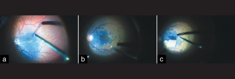
Figure 2.

Intraoperative steps of internal limiting membrane peeling (ILM) using heavy Brilliant Blue G (HBBG). (a) HBBG flows out from the injection cannula in an uninterrupted stream, sedimenting on the posterior pole. There is triamcinolone residue on the optic nerve head, from the just concluded posterior vitreous interface removal. (b) Note the excellent staining of ILM obtained with HBBG (not the same eye as in Figure a). The temporal macula did not stain uniformly due to presence of epiretinal membrane. (c) Maculorrhexis in progress: A large flap of ILM has been fashioned with end-gripping forceps
