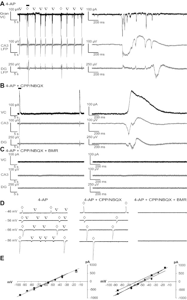
Fig. 7.

Synaptic input to DG granule neurons during distinct LFPs. A–C: example currents from whole cell voltage-clamp recordings at −60 mV with a K-gluconate-based pipette solution from a granule neuron are shown together with LFPs recorded from CA3- and DG-located pMEA electrodes. Excitatory postsynaptic current (EPSC) sequences occurring during the LFPs (A) were blocked by perfusion with ionotropic glutamate receptor (iGluR) antagonists (B), revealing large outward currents that disappeared with perfusion with BMR along with the CPP/NBQX LFP (C). The open inverted triangles indicate LFPR-CA3; the open diamonds indicate LFPP-CA3/DG. D: whole cell currents recorded from granule neurons using a pipette solution with blockers of voltage-gated Na+ and K+ channels and a higher intracellular Cl− concentration. Occurrences of LFPR-CA3 and LFPP-CA3/DG are marked by open inverted triangles and open diamonds, respectively. Whole cell currents were compared at different holding potentials in the presence and absence of blockers of iGluR and GABAA receptors. Voltages on the left of the traces are holding potentials corrected for the liquid junction potential. E: quantification of the voltage dependence of the peak current in correspondence to LFPP-CA3/DG measured from at least three events at each voltage in four distinct neurons in the absence (left) and presence (right) of CPP/NBQX.
