Fig. 1.

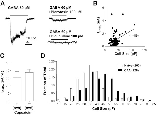
GABA current in acutely dissociated cutaneous dorsal root ganglion (DRG) neurons from adult rats. A, left: typical current evoked from a holding potential of −60 mV in response to an 8-s application of GABA (60 μM) to a small-diameter cutaneous neuron from a naive rat. Right: GABA current was almost (to 78%) completely blocked by the noncompetitive antagonist picrotoxin (100 μM) and completely blocked by the competitive antagonist bicuculline (50 μM). Scale bar on left also applies to traces on right. Data with picrotoxin were from the same neuron on left, while those with bicuculline were from another neuron with comparable baseline current. B: peak current evoked in response to 1 mM GABA (IGABA) is plotted as a function of the membrane capacitance for cutaneous neurons from naive rats. There is a small (r2 = 0.36) but significant (P < 0.01) correlation between current amplitude and membrane capacitance. C: GABA current density (current divided by membrane capacitance) is comparable (P > 0.05) even in distinct subpopulations of cutaneous neurons such as small diameter capsaicin responsive (+) and large diameter capsaicin nonresponsive (−) neurons. D: histogram of cell body size, as determined by membrane capacitance, for cutaneous neurons from naive and inflamed [Complete Freund's adjuvant (CFA)] rats illustrates the significant (P < 0.01) inflammation-induced increase in cell body size. In this and subsequent figures, data are plotted as means ± SE, where the number of neurons studied is indicated in parentheses.
