
Fig. 2.
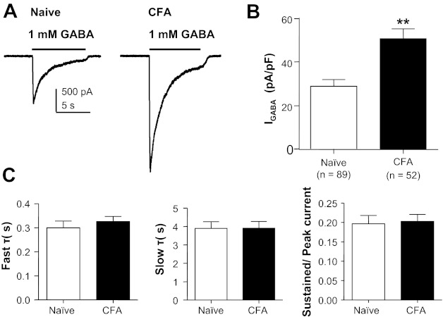
Persistent inflammation-induced increase in GABA current density. A: representative current evoked in response to 1 mM GABA applied to cutaneous neurons from a naive rat (left) and a rat 72 h after the induction of inflammation (CFA, right). B: pooled data indicated that the current density in cutaneous neurons from inflamed rats was almost double that in neurons from naive rats. C: there was no detectable difference (P > 0.05) between cutaneous neurons from naive and inflamed rats with respect to the rate of GABA current inactivation, which was defined by both fast (left) and slow (center) time constants (τ). There was also no difference (P > 0.05) between these groups with respect to the ratio of peak to sustained currents (right). **P < 0.01.