
Fig. 6.
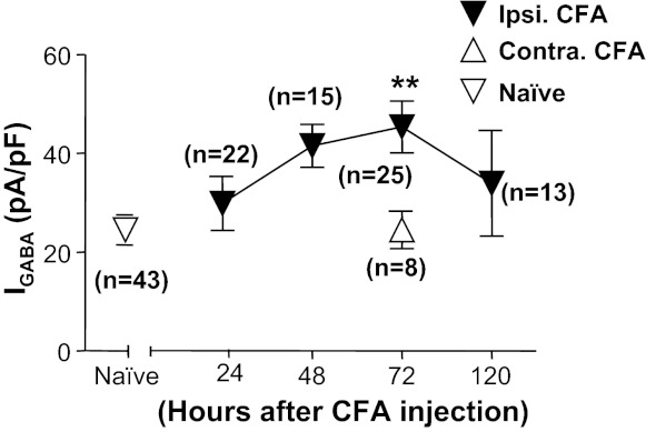
Time course of CFA-induced increase of GABA current density in cutaneous neurons. Data were collected from cutaneous neurons from naive rats and from rats 24, 48, 72, and 120 h after the induction of inflammation (ipsi CFA). A group of neurons was also studied from the side contralateral (contra) to the site of inflammation at the 72 h time point. There was a significant effect of time after inflammation relative to control, where post hoc analysis indicated that the current density at 72 h after inflammation was significantly greater than that in neurons from naive rats and in neurons contralateral to the site of inflammation. **P < 0.01.