
Fig. 9.
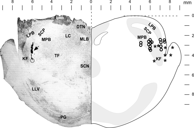
Left: section of rostral pons at the level of the caudal pole of the inferior colliculus (0 R-C) indicating the location of a microinjection in the most sensitive response area in this dog. The open circle indicates the location of the tip of the electrode at the bottom of the track (0 mm R-C, 6 mm lateral, 4.2 mm deep) that has been filled by the fluorescent beads (arrow) during injection and is just medial to KF. Right: diagram shows the most effective sites for NAL reversal of IV remi-induced bradypnea, where asterisks indicate sites based on the fluorescent bead microinjections and open circles indicate sites based on stereotaxic coordinates relative to the midline and depth for R-C coordinates from 0 to −2 mm relative to the caudal pole of the IC. NAL microinjections in and near the brachium pontis (lateral-most asterisks) resulted in nearly complete (78–100%) reversal of the remi-induced bradypnea, suggesting that μORs in the lateral parabrachial-KF complex mediate the remi-induced bradypnea. DTN, dorsal tegmental nucleus; KF, Kölliker-Fuse nucleus; LC, locus coeruleus; LLV, lateral lemniscus ventral nucleus; LPB, lateral parabrachial nucleus; MLB, medial longitudinal bundle; MPB, medial parabrachial nucleus; PG, pontine gray; SCN, superior central nucleus; SCP, superior cerebellar peduncle; TF, tegmental field.