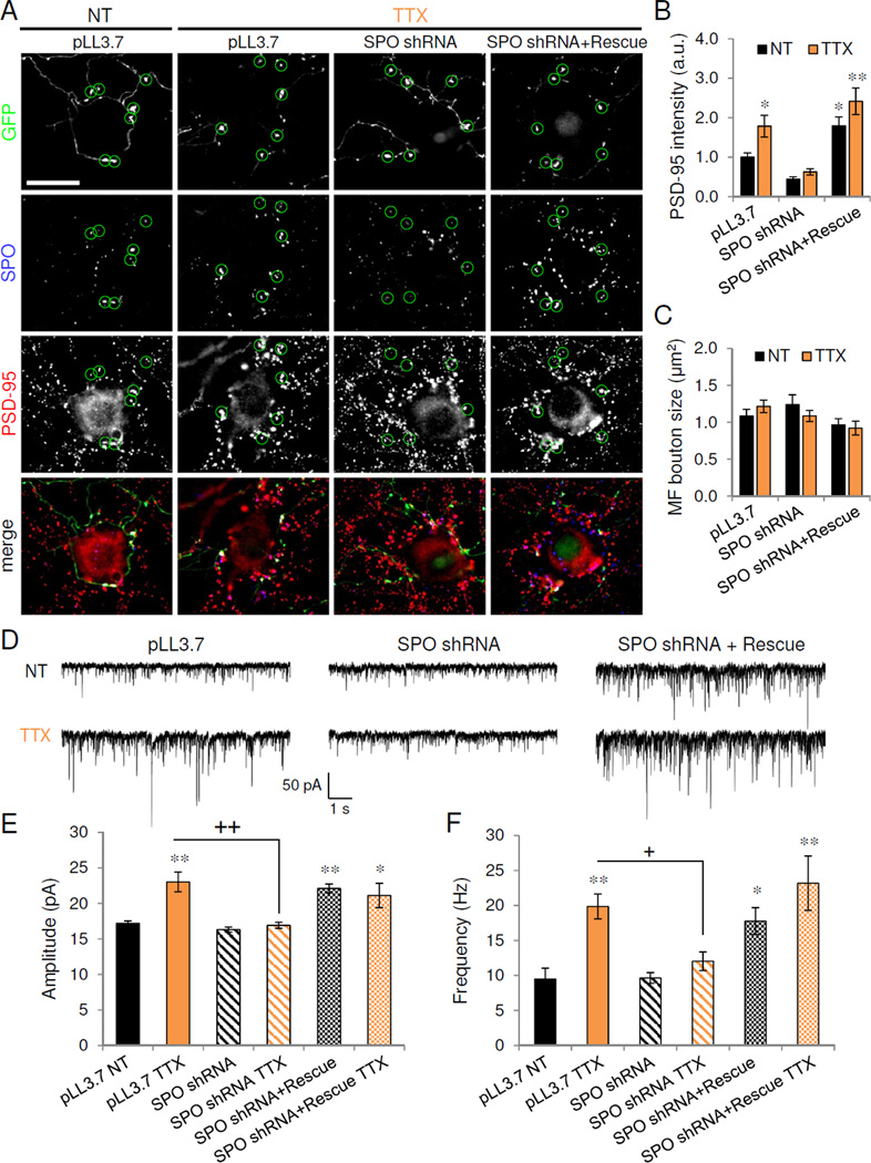
Figure 6. Presynaptic synaptoporin is required for homeostatic adaptation to inactivity.

(A) PSD-95 (red) immunolabeling of untransfected CA3 neurons receiving presynaptic innervation from synaptoporin (SPO)-positive mossy fiber terminals originating from GFP-expressing dentate granule (DG) cells transfected with vector control (pLL3.7), SPO shRNA, or SPO shRNA and SPO rescue construct. Circles indicate large GFP-positive presynaptic mossy fiber boutons. Scale, 20 µm. (B) Normalized PSD-95 puncta intensity in CA3 neurons opposing GFP-positive mossy fiber boutons transfected with vector control, SPO shRNA, or SPO shRNA+rescue construct, and treated as shown; n=12, * P<0.05, **P<0.01. (C) Mossy fiber bouton size from GFP-positive DG neurons transfected with constructs and treated as indicated. (D) Representative 10s voltage-clamp trace of AMPAR-mEPSCs recorded from untransfected targets of GFP-positive mossy fibers. DG cells were transfected with constructs indicated prior to treatment with vehicle (NT) or TTX. (E–F) Average amplitude (E) and frequency (F) of AMPAR-mEPSCs recording from untransfected control (NT) and TTX cells receiving presynaptic innervation from GFP-positive mossy fibers of DG cells transfected with constructs indicated. *P<0.05, **P<0.01 vs. NT; ns = P>0.05, + P<0.05, ++ P<0.01 vs. TTX (n=6–11). Data are means±SEM. See also Figure S6.
