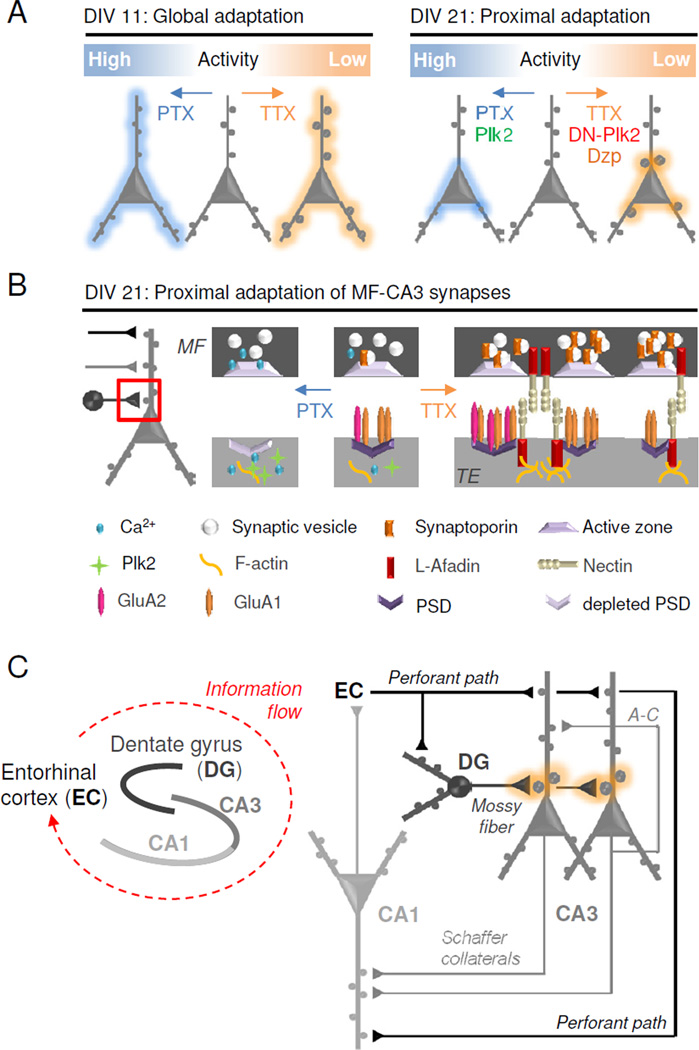
Figure 8. Proposed homeostatic gain control locus at mossy fiber-CA3 synapses.

(A) Schematic diagram of homeostatic regulation in developing (DIV 11, left) and mature (DIV 21, right) hippocampal neurons. (B) Model showing molecular mechanisms of selective synaptic adaptation between mossy fibers (MF) and TEs in mature neurons. Hyperactivity (PTX): Presynaptic Ca2+ influx leads to diminished MF expression of synaptoporin (SPO) and less vesicular release. Postsynaptic Ca2+ influx induces Plk2 expression, depletion of PSD components, and synaptic loss. Inactivity (TTX): Decreased presynaptic Ca2+ influx increases SPO expression and vesicular release in mossy fibers. Diminished postsynaptic Ca2+ influx induces l-afadin expression and burgeoning of PSDs, increasing the number of synaptic sites. GluA1 homomers may be incorporated into nascent synapses while existing synapses are strengthened by GluA1/2 heteromers. (C) Simplified hippocampal circuit diagram with MF-TE gain control (orange). Information enters the hippocampus from the entorhinal cortex (EC) via the perforant path (PP) which innervates dentate granule (DG) cells as well as distal CA3 and CA1 dendrites. Hebbian plasticity may occur in distal dendrites receiving recurrent (associational-commissural (A–C) CA3-CA3) and throughput (PP-CA3/CA1 and CA3-CA1) innervation. Homeostatic plasticity occurs at the proximal synapses of CA3 neurons which receive mossy fiber input from DG neurons.
