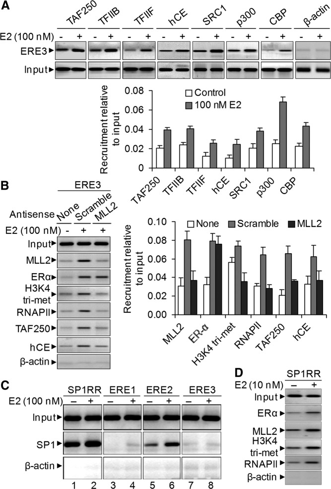
Fig. 4.

E2-induced PIC formation and recruitment of ER coactivator on the SR-B1 promoter. A, HEPG2 cells were treated with 100 nm E2 for 6 h and subjected to ChIP assay using antibodies specific to transcription factors TFIIB, TFIIF, TAF250, and hCE and ER coactivators SRC-1, p300, and CBP. The immunoprecipitated DNA fragments were PCR amplified using primers specific to the ERE3 region of SR-B1 promoter. Real-time quantification of recruitment level relative to input is shown in the bottom panel. Bars indicate se (n = 3; P ≤ 0.05). B, Roles of MLL2 on PIC assembly. HEPG2 cells were transfected with MLL2 antisense (and scramble) for 48 h and then treated with 100 nm E2 for 6 h and subjected to ChIP assay using antibodies specific to MLL2, ERα, H3K4 trimethyl, RNAPII, TAF250, and hCE. The ChIP DNA fragments were PCR amplified using primers specific to the ERE3 region of SR-B1 promoter. Real-time quantification of level of recruitment relative to input is shown in the right panel. Bars indicate se (n = 3; P ≤ 0.05). C, E2-induced recruitment of Sp1 in SR-B1 promoter. HEPG2 cells were treated with 100 nm E2 for 6 h and subjected to ChIP assay using antibodies specific to transcription factor Sp1. The immunoprecipitated DNA fragments were PCR amplified using primers specific to ERE1, ERE2, ERE3, and Sp1RR of SR-B1 promoter. D, E2-induced enrichment of ERα, MLL2, and RNAPII and E2-induced increase of H3K4 trimethylation level in Sp1RR.
