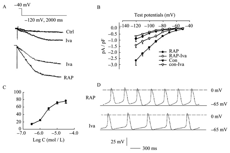
Figure 5. Effect of Ivabradine on If of PVs cardiomyocytes.

(A): peak current densities of If in RAP cells were reduced from –2.74 ± 0.52 pA/pF to -1.47 ± 0.26 pA/pF (P < 0.01, n = 12), while densities of If in control cells were reduced from –0.93 ± 0.20 pA/pF to –0.61 ± 0.05 pA/pF by 1.0 µmol/L Iva. Both inhibition effects showed significant difference (inhibition percent of 46.4% in RAP cells vs. 33.3% in control cells, P < 0.05); (B): I-V relationships demonstrated that significantly inhibited effects of 1.0 µmol/L Iva on If density more negative –90 mV potentials with a repeated-measures ANOVA; (C) Iva-induced inhibition concentration dependence of If was tested under the concentration of 0.1–10.0 µmol/L and the IC50 value was 3.2 µmol/L in RAP and control cells respectively; (D): The events of spontaneous diastolic depolarization were reduced by 1.0 µmol/L Iva. Iva: ivabradine; PVs: pulmonary vein sleeves; RAP: rapid atrial pacing.
