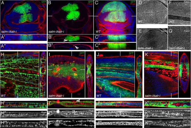
Fig. 2. Cellular effects of loss of salm and salr expression.

(A–A″) Third instar wing disc of UAS-dicer2/+; salEPv-Gal4 UAS-GFP/UAS-salm-i; UAS-salr-i/+ genotype showing the expression of GFP (green), Dlg (red) and TO-PRO (blue). A′ and A″ are transversal sections showing the three channels (A′) and the red and blue channels (A″). (B–B″) Expression of activated Cas3 (blue), GFP (green) and Phalloidin (red) in UAS-dicer2/+; salEPv-Gal4 UAS-GFP/UAS-salm-i; UAS-salr-i/+. The arrowhead in B′ indicates the localization of activated Cas3 cells that also express GFP. The arrowhead in B″ indicates the epithelial fold. (C–C″) salEPv-Gal4 UAS-GFP/+ third instar control wing disc showing the expression of GFP (green), Phalloidin (red) and To-Pro (blue). C′ and C″ are transversal sections showing the three channels (C′) and the red and green channels (C″). (D,E) Expression of FasIII in wild type (D) and UAS-dicer2/+; salEPv-Gal4 UAS-GFP/UAS-salm-i; UAS-salr-i/+ (E) third instar discs. Note the reduction in FasIII expression in discs where Sal expression is reduced. (F,G) Wild type pupal wing of 36–40 hours APF (F) and pupal wing of the same age of UAS-dicer2/+; salEPv-Gal4 UAS-GFP/UAS-salm-i; UAS-salr-i/+ genotype (G). Sal mutant cells show larger size than wild type cells, and a reduced expression of FasIII. (H–I‴) Expression of GFP (green), Phalloidin (red) and TO-PRO (blue) in UAS-dicer2/+; salEPv-Gal4 UAS-GFP/+ control pupal wings (H–H‴) and UAS-dicer2/+; salEPv-Gal4 UAS-GFP/UAS-salm-i; UAS-salr-i/+ (I–I‴) pupal wings 24–30 hours APF. H′–H‴ and I′–I‴ are tangential sections showing the expression of these three markers (H′ and I′), and the single channels with Phalloidin (H″ and I″) and TO-PRO (H‴ and I‴) expression. The arrowhead in I′ indicates extruded cells. (J–K‴) Expression of Phalloidin (red), GFP (green) and TO-PRO (blue) in UAS-dicer2/+; salEPv-Gal4 UAS-GFP/+ control pupal wings (J–J‴) and UAS-dicer2/+; salEPv-Gal4 UAS-GFP/UAS-salm-i; UAS-salr-i/+ (K–K‴) pupal wings 36–40 hours APF. Cells belonging to the sal domain are still present (labelled in green in H,I,J,K) and the epithelium show a strong phenotype of loss of integrity which is better appreciated in the sagittal sections shown in I′–I‴ and K′–K‴. Cell morphology is also strongly altered, and the wings can display indentations (I–I‴). The GFP-negative cells located between the dorsal and ventral wing surfaces, which correspond to circulating haemocytes, are marked with Phalloidin (red) in I″ and K″. White lines in B,C,H–K label the sections shown to the right and to the bottom of each panel.
