
Fig. 8. Dynamics of GFP-hFMRP within FMRP granules in HeLa cells by FRAP.
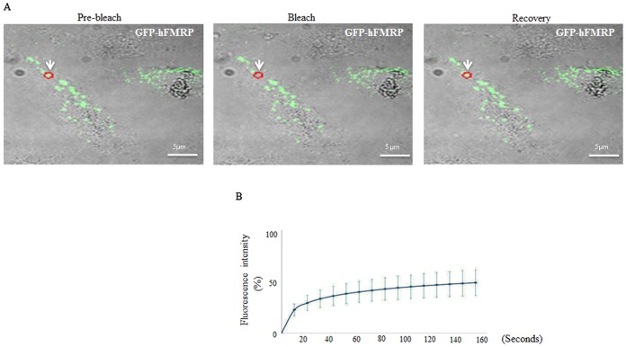
(A,B) Cells were transfected with GFP-hFMRP. Forty-eight h post transfection, a single FMRP-granule (red circle; indicated by arrow) was photobleached (A) and the fluorescence recovery (B) was recorded over 140 s using a confocal microscope as described in Fig. 7. Scale bar in A: 5 µm.