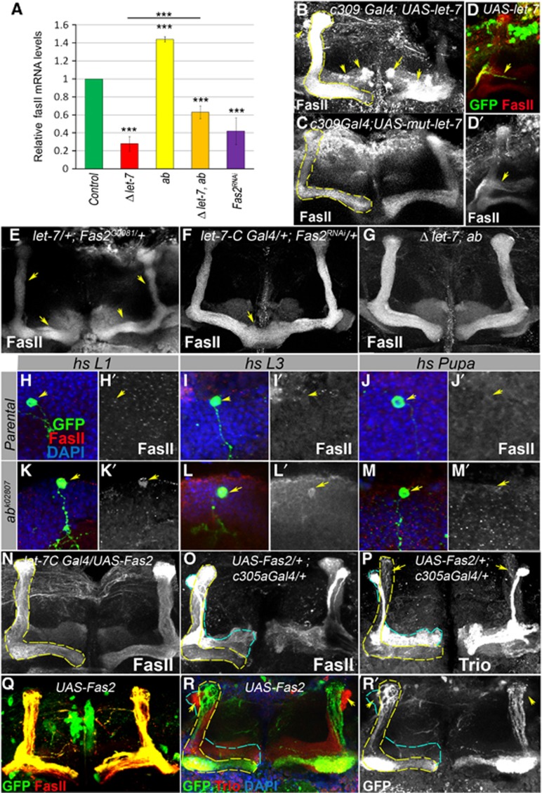
Figure 6.

Establishment of Drosophila MBs depends on the level of the cell adhesion molecule Fas II that is temporally regulated by let-7 via the transcription factor Abrupt. (A) Fas II mRNA levels are significantly decreased in the brains of let-7 mutants and increased in Ab hypomorphic mutants. Fas II decrease in let-7 mutants can be partially relieved via introduction of hypomorphic ab mutations. Downregulation of Fas II using Fas II RNAi driven by the let-7 promoter (let-7-CGK1; Fas2RNAi) results in a 60% reduction of Fas II mRNA levels (***P⩽0.001); see also Supplementary Table 9). (B) Overexpression of let-7 miRNA in all MB lobes (c309-Gal4; UAS-let-7, for the c309-Gal4 expression pattern, see Supplementary Figure S5) increases Fas II levels and induces abnormal Fas II expression in γ and α′/β′ lobes (arrows) in comparison to control (C, c309-Gal4; UAS-mut-let-7; Supplementary Table 10). (D) Single GFP marked neuron overexpressing miRNA let-7 (hsFlp; act>CD2>Gal4 UAS-GFP/UAS-let-7; L2 stage clonal induction) has an elevated level of Fas II. (E) Epistatic interaction between let-7 and Fas2 mutants results in the ‘slim α/β lobe’ phenotype (see also Supplementary Table 5). (F) Reduction of Fas II in let-7 expressing neurons results in the β-lobe fusion phenotypes with a frequency of 50%, n=12 MB lobes (see also Supplementary Table 10). (G) let-7 morphological abnormalities are rescued by reduction of Ab levels achieved using the ab amorphic and hypomorphic allelic combination in a let-7 mutant background (let-7 miR-125, ab1/let-7 miR-125, ab1D), n=12 MB lobes. (H–M) Adult MB cell body clusters containing Control (hsFlp UAS CD8GFP; tubGal80 FRT 40A/FRT 40A; tubGal4/+, H–J) and ab loss of function (hsFlp UAS CD8GFP; tubGal80 FRT 40A/abk02807FRT 40A; tubGal4/+, K–M) MARCM clones. Loss of Ab from the early-born KCs (clonal induction at the stage of L1 or L3 larva) that normally have very faint Fas II staining pattern in MB cell body clusters (H, I) caused upregulation of Fas II protein levels (K, L); higher Fas II was not observed when clones were induced in α/β lobe neurons at the 12 h APF stage (M). Arrows point to control or ab mutant clones marked with GFP (H–M) and corresponding Fas II levels (H′–M′). (N) Overexpression of Fas II with let-7-C promoter did not cause noticeable morphological changes in α/β MB lobes, while forced expression of Fas II in α′/β′ lobes (O, P) using c305a Gal4 driver (for the expression pattern, see Supplementary Figure S5) results in abnormal MB morphology resulted from projection of Trio expressing cells into α/β MB lobes. (Q, R) MBN-derived GFP marked neurons overexpressing Fas II (hsFlp; act>CD2>Gal4 UAS-GFP/UAS-Fas2; L2 stage clonal induction) did not project into α′/β′ lobes (marked with arrows, outlined with blue dashed line).
