Figure 1.

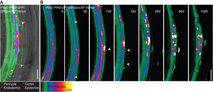
PIN3 is transiently induced in the endodermis during LRI. (A) PIN3::PIN3-GFP expression was detected in pericycle FCs (yellow arrowheads), and the overlaying endodermis cell (white arrowheads). Note that neighbouring endodermal cells are not labelled with the PIN3-GFP signal. Red arrowheads indicate the migrated nuclei expressing an enhanced DR5pro::N7:Venus auxin response reporter. A semiquantitative colour-coded heat-map of the GFP fluorescence intensity is provided. Colour-coded asterisks indicate the different root cell files. Scale bar: 25 μm. (B) Real-time analysis of PIN3::PIN3-GFP expression in the endodermis (white arrowheads) relative to FC establishment and LRI. FC establishment and LRI were followed by the accumulation of the nuclear DR5pro::N7:Venus signal (red arrowheads) in the pericycle FCs (yellow arrowheads) and the division of these nuclei. Image series depicted is a representative example from at least 10 observations, and time stated is relative to root bending. Scale bar: 20 μm.
