Figure 3.

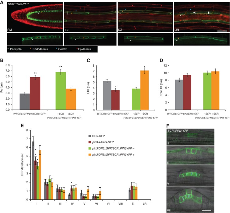
PIN3 activity in the endodermis promotes FC progression. (A) Monitoring of SCR::PIN3-YFP expression (green) in the RM, EZ, DZ and during LRI (white arrowhead). Propidium iodide (PI) counterstain (red) is shown in the upper panel. The lower panel reveals PIN3 polarity in the endodermal cells of the depicted root zones. Colour-coded asterisks indicate the different root cell files. Scale bar: 50 μm. (B, C) FC (B) and LRI (C) densities are rescued in the pin3/DR5::GFP roots expressing SCR::PIN3-YFP in the endodermis. (D) The total number of FC+LRI densities is similar to WT in all genetic backgrounds. (E) Stage distribution of LRP indicates that the decrease in stage I in pin3/DR5::GFP mutants is restored in the fluorescent-positive pin3/DR5::GFP/SCR::PIN3-YFP seedlings compared to WT/DR5::GFP. +SCR and –SCR refer to the pooled fluorescent-positive and -negative seedlings, respectively, from a segregating population in the stable pin3/DR5::GFP background (B–E). (F) The SCR promoter drives PIN3-YFP expression in the LR only from stage II onwards. Corresponding stages of LR development are indicated. Scale bar: 20 μm. Error bars represent s.e.m. (n=10–20). P-values are *<0.05, **P<0.01; Student’s t-test.
