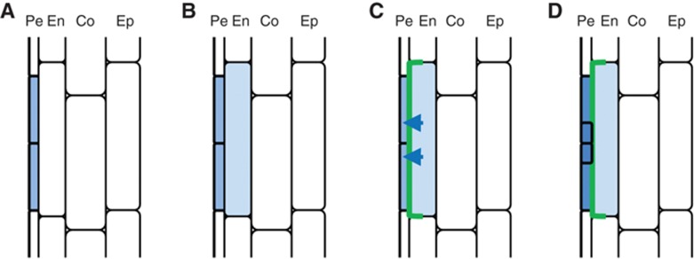
Figure 6.

Model for endodermal PIN3 regulated transition from FC to LRI. (A) Auxin response activity (blue) in a restricted number of pericycle cells indicates FC establishment. (B, C) Soon after, auxin signalling is activated in the overlaying endodermal cell (B), inducing local and transient expression of PIN3 (green) (C). In the endodermis cell, the PIN3 protein is laterally localized to the inner membrane, thereby transporting auxin towards the FCs, providing a local auxin reflux pathway. (D) This PIN3-driven auxin reflux contributes to auxin accumulation in FCs important for further progress to LRI.
