
Figure 7.
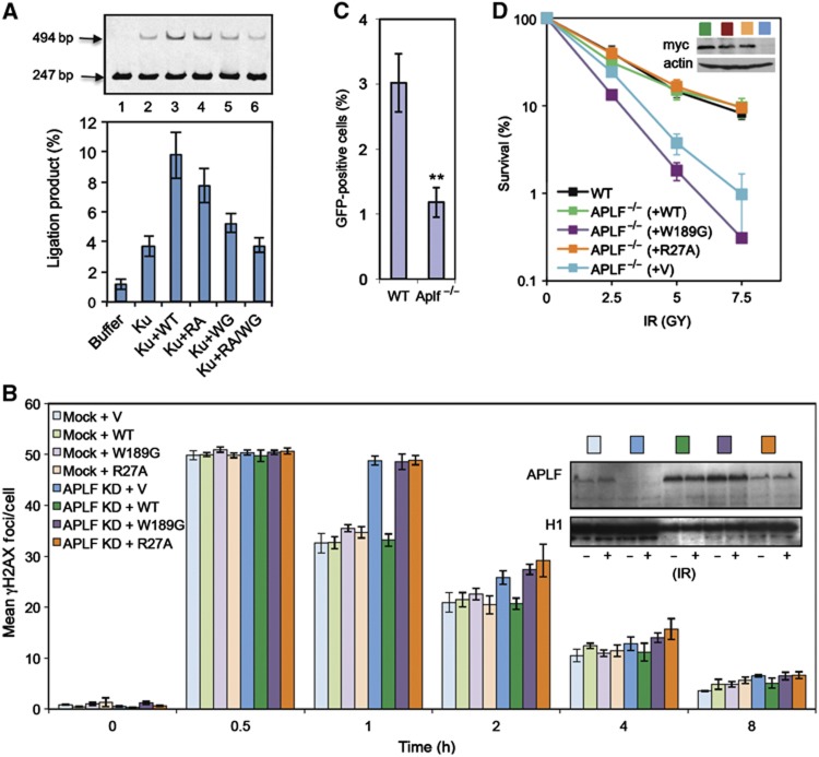
APLF scaffold activity stimulates DNA ligation in vitro and is required for rapid repair of, and resistance to, chromosomal DSBs. (A) APLF stimulates XRCC4-Lig4 activity in vitro. A Cy3-labelled 247-bp DNA duplex with one ligatable end was incubated for 30 min with recombinant XRCC4-Lig4 (10 nM) alone (‘buffer’) or in the presence or absence as indicated of recombinant Ku70/80 (‘Ku’; 20 nM), wild-type APLF (‘WT’), APLFW189G (‘WG’), APLFR27A (‘RA’), or APLFW189G/R27A (‘RA/WG’). Reaction products were fractionated by denaturing PAGE. A representative image from one experiment is presented (top), along with quantitative data from four independent experiments showing the mean (±s.e.m.) fraction of total DNA converted to a 494-bp product (bottom). (B) APLF scaffold activity is required to accelerate DSB repair. A549 cells were transiently co-transfected with empty pSUPER (‘Mock’) or pSUPER-APLF (‘APLF KD’) and either empty pcD2E expression vector (‘V’) or pcD2E encoding shRNA-resistant wild-type APLF (‘WT’), APLFR27A (‘R27A’), or APLFW189G (‘W189G’). Transfected cells were mock irradiated or γ irradiated (2 Gy) and allowed to recover for the times indicated prior to immunostaining. Data are the mean number (±s.e.m.) of γH2AX foci scored in G1 cells from three independent experiments. Inset, immunoblotting for levels of APLF and histone H1 (loading control) in untreated cells and γ-irradiated cells (30 min after irradiation). (C) Reduced plasmid re-joining in APLF−/− DT40 cells. Wild-type (Clone 18) and APLF−/− DT40 cells were co-transfected with circular RFP and linear GFP vector, 18 h before analysis by FACS. Data are the percentage of RFP-positive cells expressing GFP and are the mean (±s.e.m.) of five independent experiments. **P<0.01 by Student’s t-test. (D) Hypersensitivity of APLF−/− DT40 cells to γ radiation. Wild-type DT40 cells (clone 18), APLF−/− DT40 cells, and APLF−/− DT40 cells stably transfected with empty expression vector or with expression construct encoding myc-tagged wild-type (WT) human APLF or the indicated myc-tagged mutant human APLF were γ irradiated with the indicated dose and cell colonies counted 10 days after treatment. Inset, anti-myc Western blot depicting expression levels of the indicated recombinant APLF protein. Note that the APLF proteins expressed in these experiments were additionally tagged with an SV40 nuclear localisation signal.