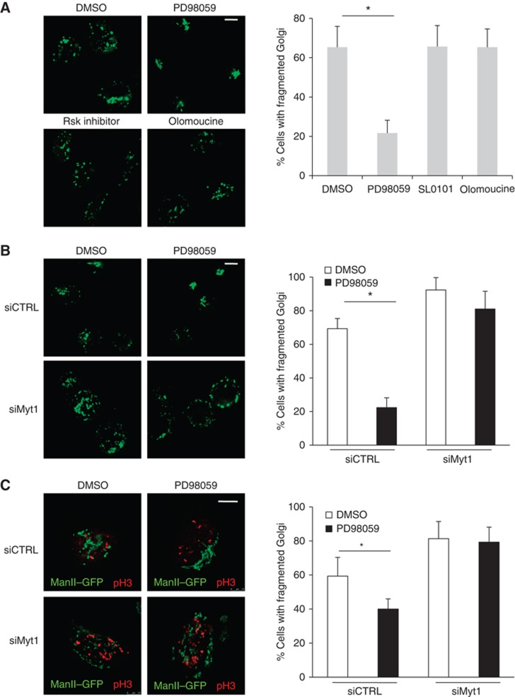
Figure 6.

MEK1 regulates fragmentation of the Golgi complex through Myt1. (A) Left panel. After incubation with thymidine for 12 h, ManII-GFP expressing HeLa cells were permeabilized, salt-washed and incubated with an ATP-regenerating system and mitotic cytosol that was pretreated with DMSO, PD, SL0101 or olomoucine. The organization of the Golgi membranes was visualized by fluorescence microscopy. Scale bar is 10 μm. Right panel. Percentage of cells with fragmented Golgi under the experimental conditions describe above. For each condition, 200 cells were counted on 2 different coverslips (mean±s.d., n=3, *P<0.05). (B) Left panel. HeLa cells stably expressing ManII-GFP were transfected with control or Myt1 specific siRNA oligo and after incubation with thymidine for 12 h, permeabilized and salt-washed cells were incubated with mitotic cytosol preincubated with DMSO or PD, and an ATP-regenerating system. The organization of the Golgi membranes was monitored by fluorescence microscopy. Scale bar is 10 μm. Right panel. Percentage of cells with fragmented Golgi under the experimental conditions describe above. For each condition, 200 cells were counted on 2 different coverslips (mean±s.d., n=3, *P<0.05). (C) Left panel. Control and Myt1 siRNA transfected HeLa cells stably expressing ManII-GFP were arrested in S-phase with a double thymidine block. Cells were washed to remove thymidine, incubated 8 h in thymidine free medium with either DMSO or PD. Then, cells were fixed and stained with an anti-phospho-histone H3 antibody and analysed by fluorescence microscopy. The images show cells in G2. Scale bar is 10 μm. Right panel. Percentage of cells with fragmented Golgi in G2-phase under the experimental conditions describe above. For each condition, 200 cells on 2 different coverslips were counted (mean±s.d., n=3, *P<0.05).
