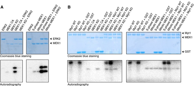
Figure 9.

Myt1 is not directly phosphorylated by MEK1 in vitro. (A) 500 ng of recombinant 6-His-tagged MEK1 proteins (constitutively active, CA; kinase dead, KD) (Left panels), and 500 ng of FLAG-tagged MEK1 proteins purified from S-phase and mitotic cells (Right panels) were incubated with 1 μg of recombinant ERK2 protein in the presence of γ-(32P)ATP for 30 min at 30°C. The reactions were stopped by the addition of SDS sample buffer and analysed by SDS/PAGE. Coomassie blue staining and the autoradiogram of the gels are shown. (B) 500 ng recombinant 6-His-tagged MEK1 proteins (CA and KD form) (Left panels) and 500 ng of FLAG-tagged MEK1 proteins purified from S-phase and mitotic cells (Right panels) were incubated with 1 μg of GST-tagged Myt1 proteins (wild type, WT; kinase dead, KD) in the presence of γ-(32P)ATP for 30 min at 30°C. The reactions were stopped by the addition of SDS sample buffer and analysed by SDS/PAGE. Coomassie blue staining and the autoradiogram of the gels are shown.
