
FIG. 3.
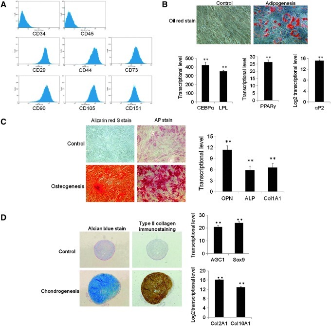
Characterization of immortalized MSCs. (A) Immortalized cells displayed surface antigen profile of MSCs. (B) Immortalized cells had adipogenic differentiation potential. Bars show the mean±SD fold change in transcript expression in MSCs from 2 patients, tested twice, relative to control MSCs in growth medium (arbitrarily set at 1). Immortalized MSCs were positive for oil red stain for lipoid deposits; adipogenic markers were uprgulated after 2 weeks of adipogenesis (**P<0.01). (C) Immortalized cells had osteogenic differentiation potential. Bars show the mean±SD fold change in transcript expression in MSCs from 2 patients, tested twice, relative to control MSCs in growth medium (arbitrarily set at 1). Immortalized MSCs were positive for alizarin red stain for calcium deposit and AP stain for alkaline phosphastase; osteogenic markers were upregulated after 2 weeks of osteogenesis (**P<0.01). (D) Immortalized cells had chondrogenic differentiation potential. Bars show the mean±SD fold change in transcript expression in MSCs from 2 patients, tested twice, relative to control MSCs in growth medium (arbitrarily set at 1). Immortalized MSCs were positive for alcian blue stain for cartilage proteoglycans and type II Collagen immunostaining for major collagen of cartilage; chondrogenic markers were upregulated after 4 weeks of chondrogenesis under pellet culture (**P<0.01).