
FIG. 14.
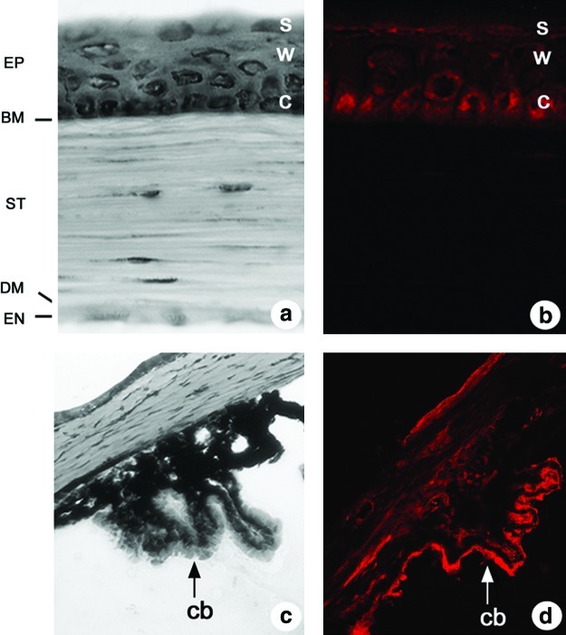
Distribution of xCT immunoreactivity in the corneal epithelium and the ciliary body. Hematoxylin-eosin stained sections of (a) the cornea and (c) the ciliary body (cb). (a) The corneal epithelium (EP) is comprised of a layer of columnar cells (c) attached to the Bowman's membrane (BM), wing cells (w), and superficial cells (s). The other layers of the cornea, from outside to inside, are the stroma (ST), the Descemet's membrane (DM), and the endothelial layer (EN). (B/D) The distribution of xCT immunoreactivity in (b) the corneal epithelium and (d) the ciliary body (To see this illustration in color the reader is referred to the web version of this article at www.liebertonline.com/ars.)