
Figure 2.
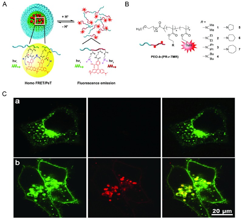
A. Design of pH-activatable micellar nanoprobes. B. Different amine groups linked to the micelle backbone to make the nanoprobes activatable at different pH upon protonation of the amine group. C. Fluorescent images of cells treated of pH-activatable nanoprobe with (top panel) or without (bottom panel) the inhibition of lysosomal acidification. Nanoprobe activation was indicated by the red fluorescence signals. Adapted from reference [30].