Figure 3.

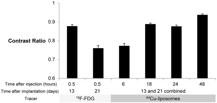
Contrast Ratio comparison with 18F-FDG and 64Cu-liposome tracers. Contrast ratio was defined as (TumorMax – MuscleMean) / (TumorMax + MuscleMean) in %ID/cc. Error bars represent SEM. For mice imaged with 18F-FDG 13 days after implantation and mice imaged 18 hours after injection with 64Cu-liposomes, n = 16 tumors, for all other groups, n = 24 tumors. Cohort imaged 13 days after implantation was maintained under anesthesia for the period between injection of 18F-FDG and imaging, while mice imaged 21 days after implantation were awakened between injection of 18F-FDG and imaging, resulting in a reduction in the contrast ratio. For the mice that remained under anesthesia during 18F-FDG image acquisition (13 days after implantation), at the earliest time point, the contrast ratio is higher for images obtained with 18F-FDG than 64Cu-liposomes, but is comparable at the 18 and 24 hour time points. At 48 hours after injection of 64Cu-liposomes, tumor contrast ratio was in all cases superior to 18F-FDG images.
