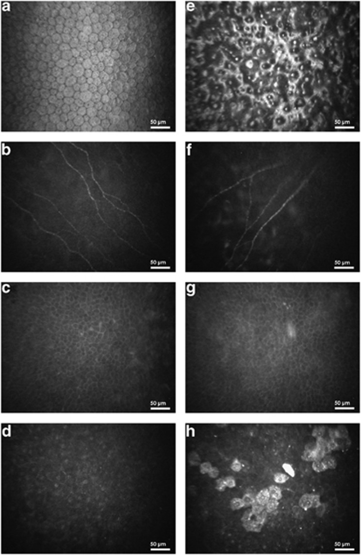
Figure 1.

Endothelial cell layer (a, e), subbasal nerve plexus (b, f), basal epithelial layer (c, g), and superficial epithelial cell layer (d, h) of one healthy control (left column) and one FECD patient (right column), respectively. ECD could not be assessed in (e).
