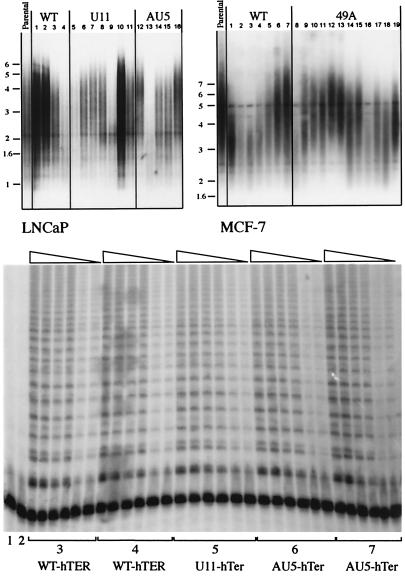
Figure 4.
MT-hTer expression does not change bulk telomere lengths or cause loss of telomerase activity. (A) Southern blotting analysis of TRF lengths. Genomic DNA was isolated 4 months after induction of expression of WT- hTER or MT-hTer expression. (Left) LNCaP clonal lines: lanes 1–4, WT-hTER expressing clonal lines R10, R11, R12, and R19; lanes 5–11, U11-hTer expressing lines 11.1, 11.3, 11.6, 11.10, 11.12, 11.14, and 11.19; lanes 12–16, AU5-hTer expressing lines 5.4, 5.5, 5.10, 5.14, and 5.15. Right: MCF-7 clonal lines: lanes 1–7, WT-hTER expressing lines F4, A4, P1, K3, K2, K1, and H6; lanes 8–19, 49A-hTer expressing lines C1, P1, M2, O1, K6, K5, K4, J2, G4, G3, D2, and D1. There was no correlation between TRF lengths and proliferation rates. (B) Telomerase activity assays (TRAP) were carried out on representative LNCaP clonal cell lines. Lane 1: lysis buffer only; lane 2: heat-inactivated WT sample; lanes 3: R11; lanes 4: R19; 5: 11.6; 6: 5.14; 7: 5.15. Each set of lanes shows 10-fold dilutions (1, 1:10 and 1:100) of each cell extract assayed; each sample was loaded in duplicate in two adjacent lanes.

