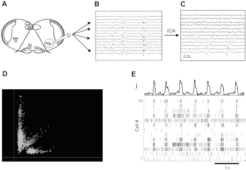
Fig. 1.

Schematic of multielectrode recording and analysis of in vitro rhythmic activity. A transverse medullary slice (A) shows the localization of the ventral respiratory group (VRG; presumptive pre-Bötzinger complex) and the placement of the twisted multiwire electrode. NA, nucleus ambiguus; IO, inferior olive; XII, hypoglossus nucleus. Multiple signals are recorded (B), and cross talk is reduced using independent component analysis (ICA; C). Extracellular spikes are detected and sorted in principal component analysis (PCA) feature space (D), identifying action potential times for individual neurons (E) in relation to the ongoing population activity (shown at top).
