Fig. 1.

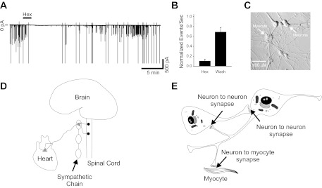
Sympathetic neurons displayed ongoing activity driven by cholinergic synaptic transmission when cultured with cardiac myocytes. A: voltage-clamp trace showing action currents (larger amplitude) and synaptic currents (smaller amplitudes) recorded at a holding potential of −60 mV. Activity was blocked by application of the nicotinic cholinergic antagonist hexamethonium (Hex; 100 μM, 2 min). B: number of synaptic events normalized to control showing that a 2-min application of 100 μM hexamethonium blocked 89.4 ± 3.7% of activity (n = 6). Action currents were excluded from excitatory postsynaptic current (EPSC) analysis. This effect was reversible, showing a 69 ± 9% recovery after 10–20 min in control saline (wash). C: phase image of a sympathetic neuron-cardiac myocyte coculture after 5 wk in culture. Neurons project widely throughout the culture dish, contacting other neurons and cardiac myocytes. D: schematic depiction of the sympathetic nervous system in vivo. Ganglionic sympathetic neurons are located in peripheral ganglia connected through the sympathetic chain along the axis of the spinal cord. Cholinergic preganglionic neurons of the spinal cord project to and innervate the sympathetic neurons, driving their activity. Sympathetic neurons form synapses within the ganglia themselves and project out to innervate tissues (such as the heart) throughout the body. E: schematic representation of our model coculture system. In culture sympathetic neurons form synapses both onto one another and onto cocultured cardiac myocytes.
