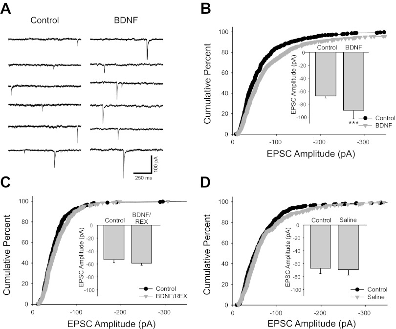
Fig. 2.
Brain-derived neurotrophic factor (BDNF) increased cholinergic EPSC amplitude. A: voltage-clamp traces recorded at −60 mV showing unitary synaptic currents recorded from a cultured sympathetic neuron. Synaptic currents were larger after 15-min application of 100 ng/ml BDNF (right). B: cumulative percent histogram plot showing a shift toward larger amplitudes of EPSCs after BDNF application (n = 11 cells; 550 and 616 EPSCs for control and BDNF, respectively; P < 0.001). The EPSC potentiation is also illustrated in a bar plot of mean amplitude (inset; −67.4 ± 3.2 pA vs. −90 ± 13 pA for control and BDNF, respectively, n = 11, ***P < 0.001). C: cumulative histogram plot showing that the p75 function-blocking antibody REX blocked the BDNF-induced EPSC enhancement (n = 5 cells; 300 and 255 EPSCs for control and BDNF/REX, respectively). EPSC amplitudes were not altered by BDNF application after incubation with the REX antibody as shown by a bar plot (inset; −53 ± 5 pA vs. −58.7 ± 3.5 pA for control and BDNF/REX conditions, respectively, n = 5). D: cumulative percent histogram plot showing no increase in EPSC amplitude after 10- to 15-min application of saline without BDNF, suggesting that the EPSC amplitude increase was not due to a time-dependent recording artifact (n = 11 cells; 517 and 550 EPSCs for control and saline, respectively, P = 0.17). No difference was seen in mean EPSC amplitudes before and after saline application as shown by a bar plot (inset; −67 ± 8 pA vs. −69 ± 8 pA for control and saline conditions, respectively, n = 11).

