Fig. 2.

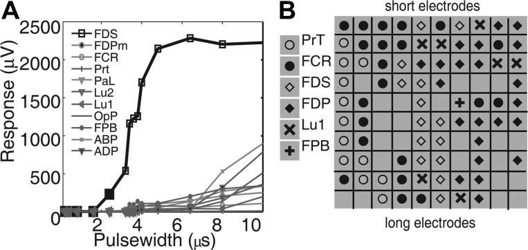
Muscle activation shows selectivity and musculotopy. A: selectivity. Stimuli of increasing pulse width evoked successively larger responses in flexor digitorum superficialis (FDS), with little or no activation of other muscles. This electrode showed a selectivity of 0.85. B: musculotopy. Each tile in the 10-by-10 grid represents an electrode on the USEA, the symbol indicating the muscle most strongly activated by that electrode. Electrodes are shown as in a cross section of the nerve with the most superficial aspect of the nerve at top. Responses in a given muscle tend to be recruited by adjacent USEA electrodes, whereas responses in other muscles are recruited by other USEA electrodes, indicating a musculotopic arrangement of nerve fibers. ABP, abductor pollicis brevis; FDPm, medial head of flexor digitorum profundus (FDP); FCR, flexor carpi radialis; PaL, palmaris longus; PrT, pronator teres; Lu1, 1st lumbrical; Lu2, 2nd lumbrical; OpP, opponens pollicis; FPB, flexor pollicis brevis; ADP, abductor pollicis brevis.
