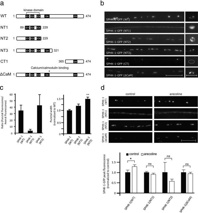
Figure 4.
Structural determinants that localize SPHK-1 at synapses and cell bodies. a, Diagram of wild-type (WT) SPHK-1-GFP and its variants used. b, Representative images of the localization of SPHK-1-GFP and indicated variants in DA/DB motor neuron axons (left) and cell body (right). c, Quantification of the ratio of punctal fluorescence to interpunctal fluorescence (left) and punctal width (right) of SPHK-1(WT)-GFP, SPHK-1(NT1)-GFP, and SPHK-1(NT3)-GFP in DA/DB motor neurons. d, Representative images (top) and quantification (bottom) of SPHK-1-GFP (WT and the indicated variants) in DA/DB motor neurons with pretreatment of control M9 (black bars) or arecoline (15 mm in M9; white bars). For all, *p < 0.05 and **p < 0.005, Student's t tests. Scale bars: 10 μm.

