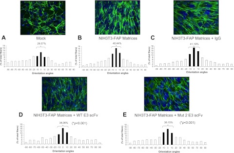
Figure 8.

Effects of anti-FAP scFvs on fibronectin organization in fibroblast-derived 3D matrices. Fibronectin fibers were analyzed by immunofluorescence in unextracted 3D matrices formed by stably transfected NIH3T3 FAP cells (B–E) or vector control (mock; A). During the formation of FAP matrices, 3 μM of mouse IgG (NIH3T3-FAP matrices + IgG; C), WT E3 scFv (NIH3T3-FAP matrices + WT E3 scFv; D) or Mut 2 E3 scFv (NIH3T3-FAP matrices + Mut 2 scFv; E) was added to the medium for 8 d. Orientation of fibronectin fibers was measured (see Materials and Methods for details). Numbers indicate the percentage of fibers positioned within 10 ° of the mode fiber orientation angle (P value between anti-FAP scFv-treated 3D matrix and nontreated control). *P < 0.05.
