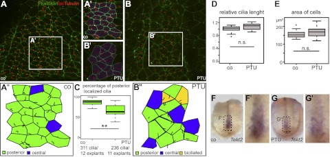
Figure 5.

Ciliation defects in the GRP of PTU-treated embryos. A–B″) Cilia and cell boundaries were visualized in dorsal explants from control (A–A″) and PTU-incubated specimens (B–B″) by immunohistochemistry (acetylated α-tubulin, red) and phalloidin staining (actin, green), respectively. C–E) Note that posterior polarization of cilia was significantly reduced (cf. quantification in C), while cilia length (D) and cell surface area (E) were unaltered. F–G′) GRP marker gene Tekt2 was markedly reduced in PTU-treated (G, G′) vs. control (F, F′) explants.
