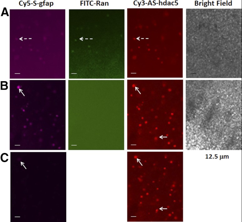
Figure 2.
Fresh living brain slices with thickness of 60 μm were harvested and incubated in F12 medium with 10% horse serum in a dish with a polylysine-free (noncoated) glass surface. A) We transfected 3 sODNs (each at 10 nM) as in Fig. 1. All sODNs were retained in neural cells (dashed arrows). After 30 min, sODN was removed by washing twice in sODN-free medium, and then the tissue was incubated in fresh medium (time 0). B, C) Photographs show different time points, 5 min (B) and 10 min (C) after sODN removal by washing in fresh medium. Solid arrows point to neural cells that exhibited slow exclusion or degradation of Cy5-S-gfap. A fast exclusion or degradation of FITC-Ran in these cells suggests the cells are viable. Scale bars = 12.5 μm.

