
Figure 2.
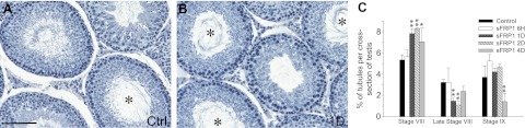
sFRP1 induces delay in spermiation. Ninety-day-old rats received 4 μg sFRP1 per testis (2.5 μg/ml, 67.6 μM; assuming a testicular volume of 1.6 ml and sFRP1 Mr at 37 kDa) in a volume of ∼200 μl at time 0 via intratesticular injection with a 28-gauge needle. Rats were terminated after 6 h and 1, 2, and 4 d with n = ∼3–5 rats/time point, including controls. Equal amount of BSA was administered to control rats. Testes were fixed in Bouin's fixative, embedded in paraffin, sectioned to 5 μm thickness using a microtome, and stained for hematoxylin for histological analysis. A, B) Testis sections from control (Ctrl; A) and 1 d after sFRP1 treatment (B). Asterisks indicate stage VIII tubules with their numbers significantly increased in testes treated with sFRP1 for 1 d. Scale bar = 60 μm. C) Numbers of stage VIII, late stage VIII, and stage IX tubules in each testis cross-section from control vs. sFRP1-treated rats were scored and represented as percentage of these staged tubules against total number of tubules in each cross-section. Late stage VIII was defined as tubules with ≥50% elongated spermatids left the tubules. Statistics were performed by comparing percentage of tubules from each stage in cross-sections of testes in rats from different treatment groups vs. rats in the control group. Results are data from n = 3 rats, with ∼800-900 tubules/testis/animal. At 1 and 2 d after sFRP1 injection, significantly more tubules were seen at stage VIII that failed to undergo spermiation. At 4 d after treatment, spermiation began to resume after sFRP1 was metabolically cleared, but the number of stage IX tubules remained significantly lower vs. control due to delay in spermiation. Bars represent means ± sd. *P < 0.05, **P < 0.01.