
Figure 4.
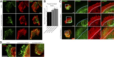
ACTN4-knockdown keratinocytes display increased focal contact area. Control iHEKs, iHEKs expressing scrambled shRNA, and ACTN4shRNA-1 cells were plated overnight onto glass coverslips and then processed for indirect immunofluorescence microscopy with antibodies against paxillin (green) together with rhodamine-conjugated phalloidin (actin, red). B) Mean ± se focal contact area was determined from images captured from the indicated cell clones and lines stained for paxillin; 3 separate experiments, 15–40 cells/experiment. *P < 0.05 vs. iHEK. C) Control iHEKs, iHEKs expressing scrambled shRNA, and ACTN4shRNA-1 cells were plated overnight onto glass coverslips and then processed for indirect immunofluorescence microscopy with antibodies against β4 integrin (green) together with rhodamine-conjugated phalloidin (actin, red). Blue line indicates base of lamellipodium. D) Same keratinocyte clones and lines were processed for indirect immunofluorescence microscopy using antibodies against paxillin (green) in combination with β4 integrin antibodies (red). Scale bars = 10 μm.