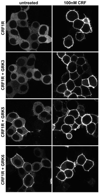
Figure 4. Recruitment of βarrestin2 by agonist-activated CRF1 receptors.
Confocal microscopy was used to evaluate the interaction of βarrestin2-GFP with CRF1 receptors (CRF1R) in real time and in live HEK293 cells co-transfected 48 h earlier with either empty vector (top panel), GRK3, GRK5 or GRK6. This representative experiment shows the distribution of βarrestin2-GFP in cells before (untreated) and after stimulation with CRF (100 nM) for 40 min. Note that both in the absence and presence of overexpressed GRKs, βarrestin2-GFP remains localized at the plasma membrane in clathrin-coated pits after translocation to cell surface CRF-activated CRF1 receptors, and does not traffic inside the cell into endocytic vesicles with internalized CRF1 receptors.

