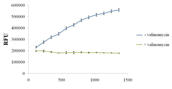
FIGURE 1.
Mitochondria staining using JC-1 stain in a Multiwell Plate Format. Mitochondria were isolated from rPCECs using the cell fractionation method and stained in a multiwell plate using the Isolated Mitochondria Staining Kit (Sigma). The upper line represents the JC-1 dye uptake of an intact mitochondrial sample. The lower line represents the dye uptake of the valinomycin treated mitochondrial control sample. RFU – Relative Fluorescence units.

