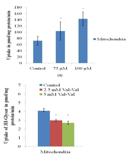
FIGURE 3.
(A) Accumulation of Rhodamine 123 (5μM) alone and in the presence of quinidine (75 and 100 μM) in the isolated mitochondria fraction from rPCECs. (B) Accumulation of [3H] Gly-sar alone and in the presence of val-val (2.5 and 5.0 mM) in the isolated mitochondria fraction from rPCECs. Values are expressed as mean ± SD (n=3). *Data were considered statistically significant for P ≤ 0.05.

