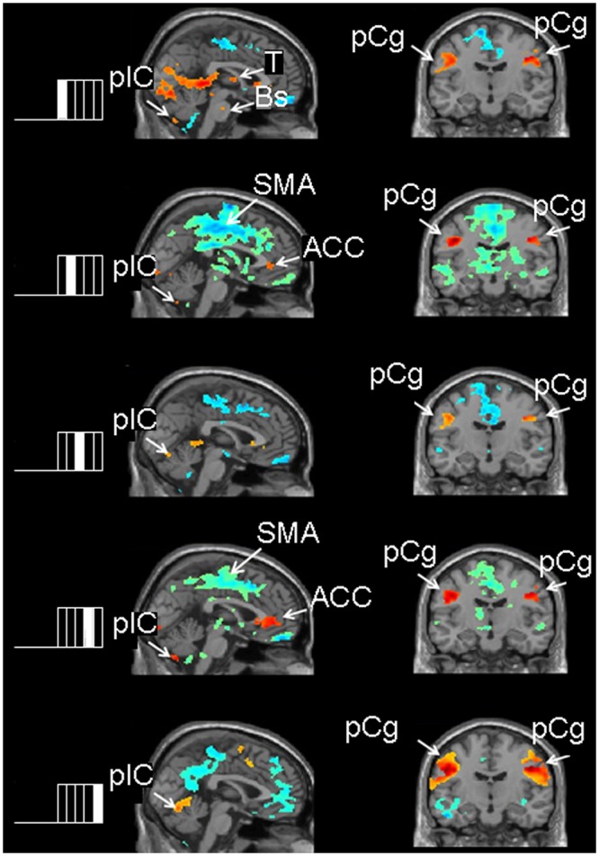
Figure 2.

Images of the contrast between each of the 5 chew-block segments relative to rest blocks. The left side of the Fig. shows the chew block partitioned into 5 segments and indicates which chew-block segment results are depicted in the adjacent brain slices. Red/yellow areas show activations or increased brain activity during the respective chewing segment compared with rest; blue/green areas show areas of decreased brain activity during the respective chewing segment compared with rest. Sagittal images show activation in the cerebellum and hypo-activations in the supplementary motor area and cingulate cortex in all 5 contrasts. Coronal images show activations in the left and right pre-central gyrus in all 5 contrasts. ACC = anterior cingulate cortex; Bs = brainstem; pCg = pre-central gyrus; plC = posterior lobe of the cerebellum, SMA = supplementary motor area, T = thalamus.
