Figure 1.
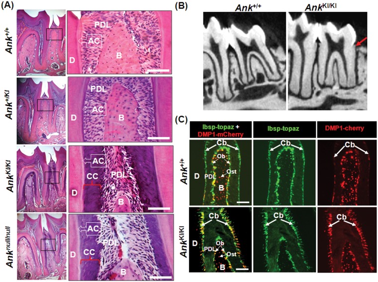
Thicker cementum deposition in AnkKI/KI mice. (A) Representative sections of 1st and 2nd molars of 10-week-old Ank+/+, Ank+/KI, AnkKI/KI, and Anknull/null mice stained with H&E. (B) Micro-CT images of 12-week-old Ank+/+ and AnkKI/KI molars. Red arrow indicates excessive cementum deposition of molar roots of AnkKI/KI mice. (C) Increased Ibsp- and Dmp1-positive cells on root surfaces of AnkKI/KI molars (4-week-old Ank+/+ and AnkKI/KI /Ibsp-Topaz/Dmp1-mCherry mice). Dentin (D); acellular cementum (AC); periodontal ligament (PDL); alveolar bone (B); cellular cementum (CC); cementoblasts (Cb); osteoblasts (Ob); osteocytes (Ost). Scale bar = 100 µm.

