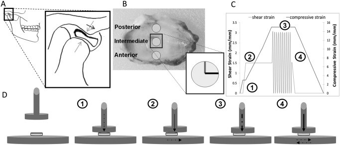
Figure 1.
Joint anatomy and simplified free-body diagram, regional sampling, and testing method. (A) The gross anatomy of the TMJ and the placement of the disc within the glenoid fossa. The insert illustrates the position of the TMJ disc when the mandibular condyle slides forward with respect to the articular eminence when the jaw opens. (B) The sampling regions of the TMJ disc and the directional notation used to orient the sample properly for testing (gray line, anteroposterior direction; black line, mediolateral direction). A representative testing strain profile is shown in (C) (description in text body), and (D) depicts the sequential testing procedure used to generate the strain profile of (C) (dashed arrows indicate active loading, and solid arrows indicate sustained loading).

