Figure 1.
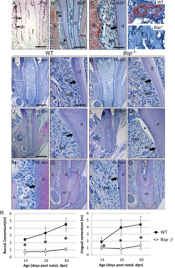
Bone sialoprotein is required for proper acellular cementum formation. (A) Bsp mRNA is expressed by cementoblasts (cb) and osteoblasts (ob) during molar tooth root development at 14 dpn. (B, C) BSP protein is localized to alveolar bone (b), acellular cementum (ac), and cellular cementum (cc) at 26 dpn. White box in B is shown at higher magnification in C to highlight acellular cementum. BSP immunolabeling in (D) WT alveolar bone is absent in (E) Bsp-/- mice. (F, G) Newly formed acellular cementum anchors periodontal ligament (pdl) attachment in WT molars. (H, I) Acellular cementum in Bsp-/- molars is thin compared with that in WT mice. In contrast to the thickened acellular cementum in (J, K) WT mice at 26 dpn, (L, M) Bsp-/- molars feature stunted acellular cementum, and regions of PDL detachment from the tooth (*) were common, indicating defects in the cementum-PDL interface. The well-organized attachment of the acellular cementum, PDL, and bone in (N) WT molars at 60 dpn is severely degraded in (P) Bsp-/- molars, where stunted acellular cementum is associated with PDL disorganization. In contrast, cellular cementum in (O) WT and (Q) Bsp-/- mice at 60 dpn is not different in size. (R) Both buccal and lingual acellular cementum samples are significantly reduced in thickness in Bsp-/- mice compared with their WT counterparts (*indicates significant difference, p < 0.05). Scale bar is 200 µm in panels A, B, F, H, O, and Q (original, 100X), 50 µm in panels C, D, E, G, I, K, M, N, and P (original, 400X), and 400 µm in panels J and L (original, 50X).

