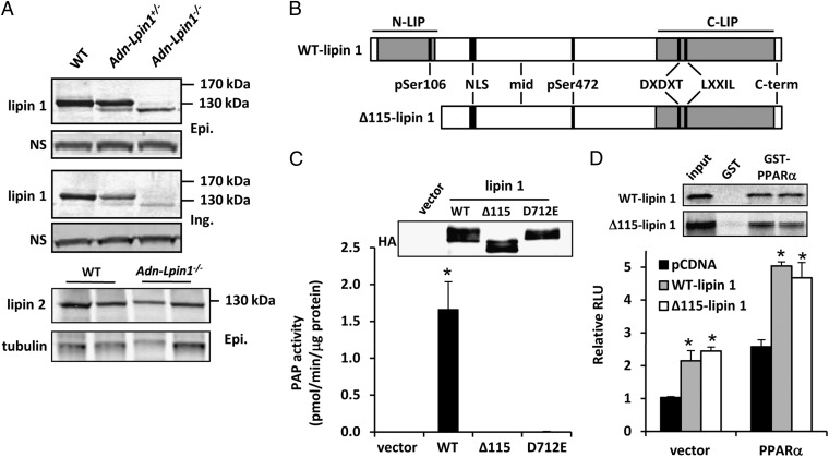
Fig. 1.
A truncated lipin 1 protein is expressed in Adn-Lpin1−/− mice. (A) Western blot analysis for lipin 1 and lipin 2 in WAT [epididymal (Epi.) and inguinal (Ing.)] of 6- to 8-wk-old male WT, Adn-Lpin1+/−, and Adn-Lpin1−/− mice. (B) Schematic depicting the domain structure of full-length and Δ115-lipin 1. The approximate binding locations of the antibodies used in Fig. S1 are shown. DXDXT, PAP catalytic site; LXXIL, nuclear receptor interaction domain; NLS, nuclear localization sequence/polybasic domain; N-LIP, N-terminus lipin homology domain; C-LIP, C-terminus lipin homology domain. (C) Graph showing Mg2+-dependent PAP activity of the indicated proteins overexpressed in 293 cells and immunopurified with HA antibody. Western blots to detect overexpressed protein are shown. (D) Graph showing luciferase activity in transfection studies using the PPAR-responsive acyl CoA oxidase-thymidine kinase luciferase reporter construct and the indicated expression constructs. GST-pull down images are shown above graph.

