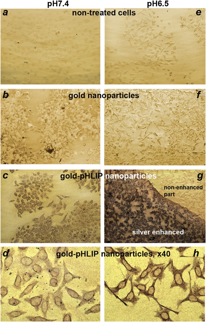
Fig. 2.
Cellular uptake of gold-pHLIP and gold nanoparticles. Nanoparticles (2 μM, 200 μL) were incubated with HeLa-GFP cells at pH 7.4 or 6.5. After 1 h, cells were washed, fixed, and stained with silver enhancement solution, resulting in the deposition of silver on gold nanoparticles to form micrometer-sized particles, which were visualized under a light microscope The images A–G and D–H were taken with 10× and 40× objectives, respectively.

