Fig. 3.
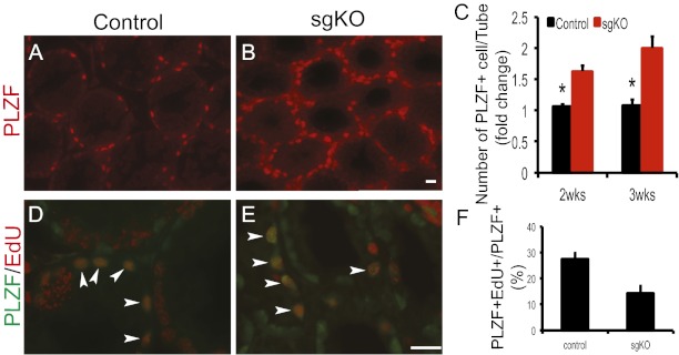
Accumulation of PLZF-positive spermatogonia in Rdh10 sgKO mutant testes. (A and B) Immunofluorescence staining for PLZF (red) in sections of 2-wk-old control (A) and Rdh10 sgKO (B) testes. (Scale bar: 20 μm.) (C) Quantification of PLZF-positive cells per seminiferous tubule in 2-wk-old testes. (D and E) Immunostaining for PLZF (green) and EdU (red) in sections of 2-wk-old control (D) and Rdh10 sgKO (E) testes. Arrowheads indicate both PLZF- and EdU-positive spermatogonia (orange). (Scale bar: 20 μm.) (F) Quantification of proliferative spermatogonia in control and Rdh10 sgKO testes at age 2 wk. The number of EdU-positive cells per number of PLZF-positive cells was recorded. All seminiferous tubules at each section were counted (n = 3–5). Error bars represent SD. *P < 0.05, Student t test.

