Fig. 4.
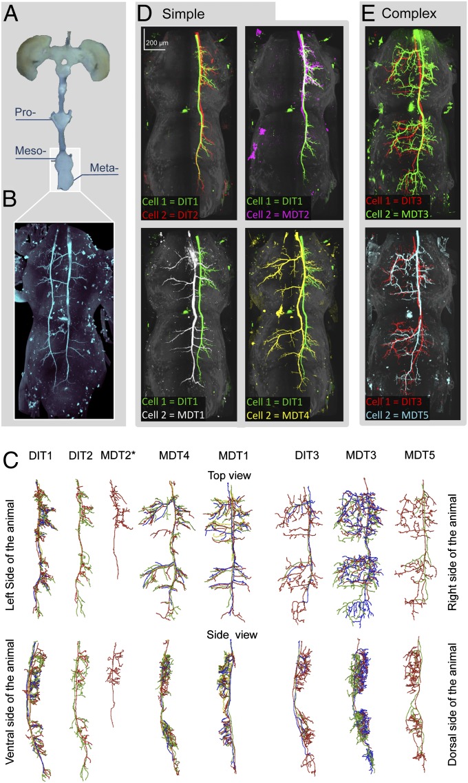
In the wing motor centers, all TSDNs share morphological features. (A) The mesothoracic, metathoracic, and first abdominal ganglia were imaged and warped to allow morphological comparison. (B) Unilateral DIT2 on the left and bilateral MDT1 on the right, injected in the same animal, target the same locations. (C) Traces within each TSDN type (grouped according to the electrophysiological results) are consistent, so the most complete fill from each TSDN type was used for comparison. TSDNs were categorized into “simple” or “complex” cells. A pairwise comparison (dorsal views) shows that unilateral simple cells, DIT1 (green), DIT2 (red), and MDT2 (magenta), are indistinguishable from each other (D, Upper), but the bilateral simple cells MDT1 (white) and MDT4 (yellow) display specific branching patterns (D, Lower). However, all simple TSDNs target the same location. In contrast, pairwise comparisons between the complex cells, DIT3 (red), MDT3 (green), and MDT5 (cyan) (E, all panels) are less informative because their additional intricate branching exhibits higher variability, particularly in the medial region of the ganglia (traces in C).

