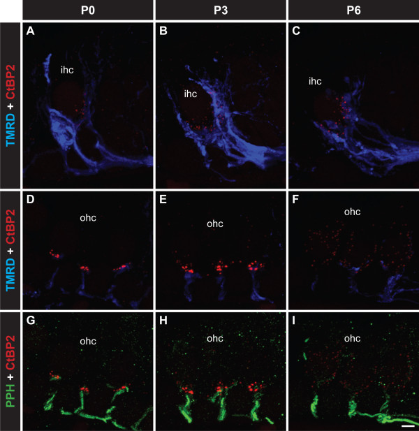
Figure 1.
Visualization of synaptic ribbons during the development of the innervation of IHCs and OHCs by type I and type II afferent fibres respectively. All images are from the mid-turn of the cochlea. Triple labelling showing CtBP2/RIBEYE puncta to mark synaptic ribbons (red), type I nerve fibres labelled with TMRD (blue; A-F) and type II fibres labelled with anti-peripherin (green; G-I) from P0–P6. A-C TMRD-positive type I fibres innervating the IHCs, initially extending to the apical cell regions before consolidating at the basolateral region where the synaptic ribbons and localised. D-F TMRD-positive type I fibres also temporarily innervate the OHCs, localising at the basal region of the OHCs where synaptic ribbons are localised. At P6, the ribbons disperse in parallel with retraction of type I fibres. G-I Peripherin-positive type II fibres innervating the OHCs. The formation of the outer spiral bundles proceeds despite dispersal of the presynaptic ribbons. Scale bar 5 mm.

