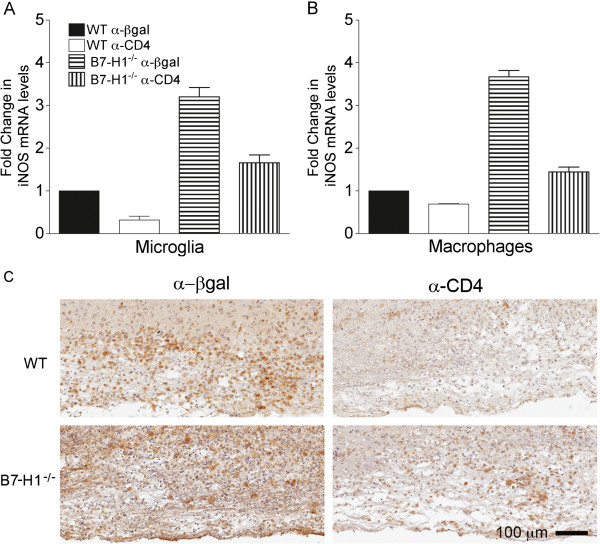
Figure 10.
Absence of CD4 T cells reduces microglia/macrophage activation. Wild-type (WT) and B7-H1−/− mice were treated with α-CD4 or control α-βgalactosidase (α-βgal) monoclonal antibody. (A,B) Fluorescence-activated cell sorting-purified CD45lo microglia and CD45hiF4/80+ macrophages from pooled brains (n = 6 to 8) were assessed for transcript levels of inducible nitric oxide synthase (iNOS) relative to GAPDH × 1000 at 10 days post-infection (p.i). Transcript levels are presented as the fold change relative to levels from WT samples set to 1. Data depict the mean ± SEM of two independent experiments. (C) Microglia/macrophage activation assessed at day 10 p.i. by Mac-3 staining of spinal cords from WT (upper panels) and B7-H1−/− (lower panels) mice that were depleted or sufficient in CD4 T cells.

