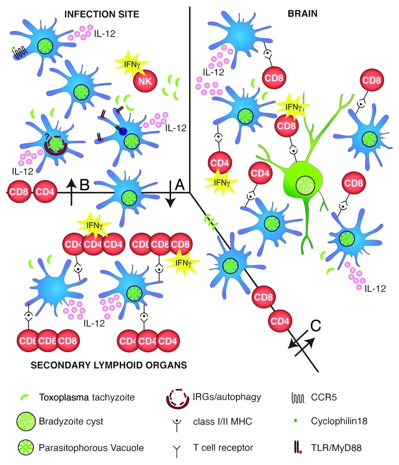
Figure 1. Complexity of dendritic cells interactions with Toxoplasma gondii on its way to achieve persistence in the host. Infection site: Toxoplasma enters an intermediate host’s body either via the natural route of infection in the gut or as in numerous studies covered here, after being intraperitoneally injected as tachyzoites. Regardless, dendritic cells (DCs) present the first line of defense. Toxoplasma infected and bystander DCs secrete the cytokine IL-12, which in turn stimulates the production of IFNγ by natural killer (NK) cells. Molecular recognition of Toxoplasma products by DCs proceeds via CCR5 sensing Toxoplasma cyclophilin18, TLR-mediated sensing of Toxoplasma profilin or other yet unknown parasite products. It remains to be investigated if IFNγ-upregulated GTPases and autophagy contribute to parasite elimination in DCs as already shown in macrophages. Arrow (A): Infected and activated bystander DCs travel from the infection site to the secondary lymphoid organ. Secondary lymphoid organ: Infected and activated bystander DCs produce IL-12 and activate CD4+ and CD8+ T cells to proliferate and produce IFNγ to activate effector molecules and mechanisms. Arrow (B): Proliferated and activated CD4+ and CD8+ T cells travel back to the infection site. Arrow (C): Infected DCs/monocytes are used as a Trojan horse by Toxoplasma to cross the blood brain barrier. Next activated CD4+ and CD8+ T cells cross the blood-brain barrier. Brain: Toxoplasma-infected and bystander DCs, astrocytes and microglia can present antigens to CD4+ and CD8+ T cells which secrete IFNγ. IFNγ secretion by CD8+ T cells is the dominant and necessary immune response for the parasite to be maintained in the bradyzoite stage and to avoid recrudescence to tachyzoites.

An official website of the United States government
Here's how you know
Official websites use .gov
A
.gov website belongs to an official
government organization in the United States.
Secure .gov websites use HTTPS
A lock (
) or https:// means you've safely
connected to the .gov website. Share sensitive
information only on official, secure websites.
